2023: Volume 5, Issue 1
Past Issues
 Abstract
  PDF
PDFThe Quality of Periapical Dental X-Ray in Wad Medani Dental Teaching Hospital, Sudan
Ahmed Esam1, Alfadel Ameer A2,*
1Faculty of Dentistry UOG, University of Gezira, Sudan
2Faculty of Dentistry UOG, University of Gezira, Sudan
*Corresponding author: Alfadel Ameer, BDS, Faculty of Dentistry UOG, University of Gezira, Sudan; Email: [email protected]
Received Date: March 6, 2023
Publication Date: April 17, 2023
Citation: Esam A, et al. (2023). The Quality of Periapical Dental X-Ray in Wad Medani Dental Teaching Hospital, Sudan. Dental. 5(1):10.
Copyright: Esam A, et al. © (2023).
ABSTRACT
Quality of x-ray is one of important dental investigation and help to Good Diagnostic Quality “Radiographs are necessary for the evaluation and diagnosis of many oral conditions and diseases. Radiographs should be specific to the needs and requirements of each particular patien”, low quality is effects in diagnosis and treatment. The aim of this study is to assess the quality of X ray in Wad Medani Dantal teaching hospital and its impact on work.
The result:
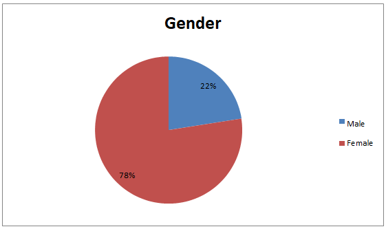
- Among group was selected, 22.8% were male where as 77.2%were female.
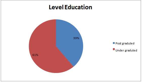
- The educational level: under graduate 62.2% , post graduate 37.8%.
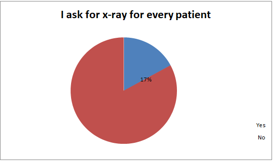
- The dentist asks for X-ray for every patient about 16.5%.
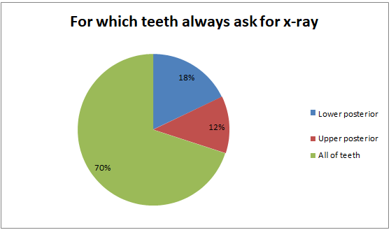
- The most dentists ask for X-ray for endodontic 52%.
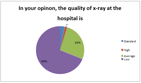
- The quality of X-ray at the hospital is low by 69, 3% .
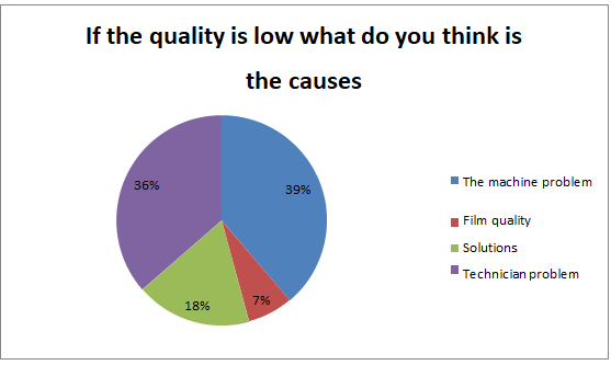
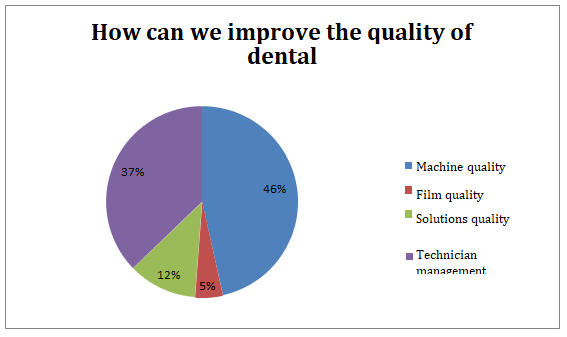
- The cause of the low quality is : the machine quality, 37.8% and the technician problem, 37%.
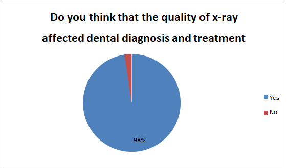
- The quality of X-ray is affects dental diagnosis and treatment about 97.6%
INTRODUCTION
Dental X-rays (radiographs) are images of your teeth that your dentist uses to evaluate your oral health. These X-rays are used with low levels of radiation to capture images of the interior of your teeth and gums. This can help your dentist to identify problems, like cavities, tooth decay, and impacted teeth.
Dental X-rays may seem complex, but they’re actually very common tools that are just as important as your teeth cleanings.
Dental X-rays are typically performed yearly. They can happen more often if your dentist is tracking the progress of a dental problem or treatment. Like brushing and flossing, getting regular dental X-rays is an integral part of your overall oral healths.
Having a good checkup can be a relief, but this doesn’t mean you shouldn’t keep getting X-rays. Depending on your age, health, and insurance coverage, X-rays may be performed every one to two years. Be sure to commit to your appointments and see your dentist sooner if you experience any pain or other changes in your mouth.
There are two main types of dental X-rays: intraoral (meaning the X-ray film is inside the mouth) and extraoral (meaning the X-ray film is outside the mouth).
Intraoral X-rays are the most common type of dental X-ray taken. These X-rays provide a lot of detail and allow your dentist to find cavities, check the health of the tooth root and bone surrounding the tooth, check the status of developing teeth, and monitor the general health of your teeth and jawbone.
Extraoral X-rays show teeth, but their main focus is the jaw and skull. These X-rays do not provide the detail found with intraoral X-rays and therefore are not used for detecting cavities or for identifying problems with individual teeth. Instead, extraoral X-rays are used to look for impacted teeth, monitor growth and development of the jaws in relation to the teeth, and to identify potential problems between teeth and jaws and the temporomandibular joint (TMJ, see temporomandibular disorders for more information) or other bones of the face.
The quality of radiographic images depends on how the image was taken, what image receptor was used, and how the visual image was created. The high quality of dental x-ray helps to provide good diagnostic quality, consistent results, lowest possible dose to patient, and cost effective.
The effects of poor radiographic technique are the same whatever type of image receptor is used. These technique errors have already been covered in detail in relation to the three main projections used in dentistry, namely periapicals, bitewings and panoramic radiographs.
Practical factors influencing film-based image quality
In practical terms, the various factors that can influence overall film-captured image quality can be divided into factors related to The X-ray equipment, The image receptor The patient , The operator and radiographic technique.
Overexposure is caused by faulty X-ray equipment, incorrect exposure time setting, and overdevelopment due to excessive time in the developer solution.
Fogging is caused by poor storage conditions, old film stock, faulty cassettes, faulty darkroom/processing units, unsafe safe-light, but underexposure lead to incorrect exposure time setting. Also underdevelopment may be due to inadequate time in the developer solution, and too cold developer solution.
Justification
I choose to study the issue of X-ray quality for a variety of reasons, including: To assess the application of quality assurance standards for digital dental radiography in everyday clinical practice. Fiercely contested is the radiograph clinically necessary is there a net benefit from taking a radiograph Does the clinical examination warrant the x-ray.
Does the background or symptoms call for an x-ray?
Objectives
General objective:-
The aim of this study is to assess the quality of X ray in Wad Medani Dental teaching hospital and its impact on work.
Study design
Methodology
This study was a descriptive analytic cross sectional type held at Wad Madani Dental teaching hospital.
Data collection and tools
The study used a cross-sectional descriptive survey design in the form of a questionnaire of 11 questions about: (demographic data, evaluation of dental x-ray in hospital, quality of dental radiograph, related b/w low quality of x-ray and misdiagnosis, prevalence of most radiograph tooth).
Site
Various places in Wad Madani Dental teaching hospital and randomly selected.
Target Population
Dentist in Wad Madani Dental teaching hospital.
Sampling and Sample Frame
Simple random sampling was used to select my sample.
Selection Criteria
Inclusion criteria: Dentist in Wad Medani Dental hospital between under graduate and post graduate who agree to participate in the study.
Exclusion Criteria
No exclusion in may sample
Ethical Considerations
All study participants gave informed consent prior to participating in the study which was conducted on an anonymous basis.
Data Analysis
Data was been analysis using manual method.
Literature Review
- Dental practitioners in the United States are exposing more than 400 million dental radiographs per year. Major national studies have indicated that improvements in radiographic quality are needed. Quality assurance (QA) programs used in medical radiology are designed to produce radiographs that are of high quality, use the least amount of radiation, and are produced at minimum cost. Preliminary recommendations are presented from the Quality Assurance Committee of the American Academy of Dental Radiology as an outline for establishing preventive maintenance of x-ray systems and a preliminary method for determining appropriate QA monitoring levels for dentistry. Included are recommendations for three stages of dental radiology QA. These are preliminary ratings and are expected to change as more dental QA information becomes available. “The American Academy of Dental Radiology Quality Assurance Committee 15 May 2005”.
- An adequate quality radiograph is one, which provides the required diagnostic information. However the quality of radiograph depends upon several contributory factors. Where the practioners is in any doubt about the reasons for poor radiographic quality It is helpful to systematically target the problem areas. This is achieved by carrying out a film reject analysis. “Shams Ul Nisa (Author)2016”
- An organised effort by the staff operating a facility to ensure that the diagnostic images produced by the facility are of a sufficiently high quality so that they consistently provide diagnostic information at the lowest possible cost and with the least possible exposure of the patient to radiation. “WHO 2005”.
- Radiation dosimetry for determining the optimum image quality with the lowest radiation exposure to the patient was carried out. The best image quality with the lowest exposure dose was assessed for conventional intraoral X-ray film (Kodak type E) and the digital processing sensor (RVG 5200). Radiation survey level was done during this study for safety and protection Purposes. “King Abdul Aziz University (KAU) Dental Hospital”.
- Quality assurance programmes for dental radiography are needed to ensure that images are consistently of a high standard. All aspects of the imaging process must be monitored to reduce the number of repeat radiographs needed, and to ensure radiography is carried out efficiently. ” Jane Luker 2014”.
- The purpose of Quality Assurance (QA) in dental radiology is to ensure consistently adequate diagnostic information, while radiation doses are controlled to be as low as reasonably achievable. “1996. European Guidelines on Quality criteria”.
RESULTS OF DATA ANALYSIS
RESULTS
The study sample consist of post graduate 38%, under graduate 61%, 22.5% were Males and 77.5% were Females.
Most dentists are not asking for x-ray for every patient (82.9%).
Most ask for x-ray in endodontic reason (51.2%) to the all teeth (69%).
The quality of x-ray is low (69%).
In this sample, (97.7%) low x-ray quality effect of diagnosis and treatment.
In this sample the main cause of low quality is machine problems (38.8%).
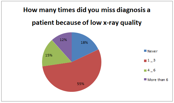
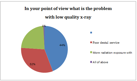
In this sample the main problem with low quality of x-ray is misleading diagnosis (44.2%).
DISCUSSION
This study revealed that assessment of x-ray quality Wad Medani Dental teaching hospital assist the quality in study area was 69% low and 26.4% average which is higher than the most previous study.
This difference may be attributed to the difference in quality of x-ray , methodology , misdiagnostic effects as well as failure RCT and difficult extracting lower 8.
As a result of all these variables, film faults and alterations in image quality are inevitable. However, since the diagnostic yield from radiography is related directly to the quality of the image, regular checks and monitoring of these variables are essential to achieve and improve the quality of radiographs. It is these checks which form the basis of quality assurance programmes.
CONCLUSION
In Wad Medani Dental teaching hospital the quality of x-ray is low (69%), the main causes is machine quality problem (38.8%) and technician problem (36.4%).
RECOMMENDATION
-Improve the machine quality and using with profession technician.
-Improve the image and solution quality.
-The technician must respect and adhere to the working hours.
-There is an increased demand to start educational programs for dental student and dentist and teachers to increased their dental knowledge and management about radiograph machine.
-Select the high quality of solutions and good type of film, using the criteria given for film quality.
-As a minimum target, no greater than 20% of radiographs should be of unacceptable quality.
-As soon as possible, insert the digital x-ray machine
REFERENCES
- Parks ET, Williamson GF. (2002). Digital radiography: an overview. J Contemp Dent Pract. 3:23–39.
- van der Stelt PF. (2005). Filmless imaging. The uses of digital radiography in dental practice. JADA. 136(10):1379-1387.
- Parks ET. (2008). Digital radiographic imaging. Is the dental practice ready? JADA. 139:447–481
- Conover GL, Hildebolt CF, Anthony D. (1995). Objective and subjective evaluations of Kodak Ekta-speed Plus dental x-ray film. Oral Surg Oral Med Oral Pathol Oral Radiol Endod. 79:246–250.
- Svenson B, Welander U, Shi XQ, Stamatakis H, Tronje G. (1997). A sensitometric comparison of four dental X-ray films and their diagnostic accuracy. Dentomaxillofac Radiol. 26(4):230–235.
- E.M. Tjelmeland, W.S. Moore, C.B. Hermesch, D.J. Buikema. (1998). A perceptibility curve comparison of Ultra-speed and Ektaspeed Plus films.” Oral Surg Oral Med Oral Pathol Oral Radiol Endod 85:488–485.
- Syriopoulos K, Velders XL, Sanderink GC, van Der Stelt PF. (2001). Sensitometric and clinical evalu-ation of a new F-speed dental X-ray film. Dentomaxillofac Radiol 30:40–44.
- Bernstein DI, Clark SJ, Scheetz JP, Farman AG, Rosenson B. (2003). Perceived quality of radio-graphic images after rapid processing of D- and F-speed direct-exposure intraoral x- ray films. Oral Surg Oral Med Oral Pathol Oral Radiol. 96:486–491.
- National Council on Radiation Protection and Measurements, NCRP report 145: Radiation Protec-tion in Dentistry. National Council on Radiation Protection and Measurements, Bethesda, MD,2003.
- American Dental Association Council on Scientific Affairs. “The use of dental radiographs: Updateand recommendations American Dental Association Council on Scientific Affairs.” JADA 137.
- National Council on Radiation Protection and Measurements. NCRP Report 172: Reference Levels and Achievable Doses in Medical and Dental Imaging: Recommendations for the United States.
- National Council on Radiation Protection and Measurements, Bethesda, MD, 2012.