2026: Volume 8, Issue 1
Current Issue
 Abstract
  PDF
PDFComprehensive Oral Rehabilitation of Mixed Dentition Affected by Amelogenesis Imperfecta: A Clinical Case Report
Farah Chouchene 1,3*, Fatma Masmoudi 2,3, Ahlem Baaziz 2,3
1Assistant Professor, Pédiatric Dentistry, Dentistry Department Ibn El Jazzar University Hospital, Kairouan, Faculty of Dental Medicine of Monastir, Tunisia
2Professor in Pediatric Dentistry, Pediatric and Preventive Dentistry Department, Faculty of Dental Medicine of Monastir, Tunisia
3Laboratory of Biological, Clinical and Dento-Facial Approach, University of Monastir, Monastir, Tunisia
*Correspondent author: Farah Chouchene, Assistant Professor, Pediatric Dentistry, Dentistry Department Ibn El Jazzar University Hospital, Kairouan, Faculty of Dental Medicine of Monastir, Tunisia, Email : [email protected]
Received: July 30, 2026
Published: August 24, 2026
Citation: Chouchene F, et al. (2026). Comprehensive Oral Rehabilitation of Mixed Dentition Affected by Amelogenesis Imperfecta: A Clinical Case Report. Dental. 8(1):24.
Copyright: Chouchene F, et al. © (2026).
ABSTRACT
Background: Amelogenesis imperfecta (AI) is a group of rare inherited enamel defects characterized by qualitative and/or quantitative abnormalities of enamel formation. Children with AI frequently experience tooth hypersensitivity, rapid enamel loss, compromised mastication, and esthetic concerns that may adversely affect their oral health-related quality of life.
Case Presentation: This report describes the multidisciplinary management of a 10-year-old girl with generalized amelogenesis imperfecta presenting with extensive enamel breakdown, yellow-brown discoloration, dentin exposure, severe hypersensitivity, and compromised esthetics. The clinical phenotype was considered predominantly hypocalcified based on the extensive post-eruptive enamel breakdown and qualitative enamel defects. The patient also presented with an Angle Class II Division 1 dental relationship, increased overjet, and anterior deep overbite. The treatment objectives were to preserve dental tissues, relieve hypersensitivity, restore masticatory function, improve esthetics, and optimize the developing occlusion.
Results: Immediate improvement in dental esthetics, elimination of hypersensitivity, restoration of masticatory efficiency, and increased patient satisfaction were achieved. At 18 months follow-up, the restorations remained clinically satisfactory, with good restoration integrity, maintained occlusal rehabilitation, and satisfactory oral hygiene.
Conclusion: Early multidisciplinary rehabilitation using conservative full-coverage restorations and individualized interceptive orthodontic management can provide effective functional and esthetic rehabilitation in children with AI during the mixed dentition stage. Regular long-term follow-up remains essential to monitor restoration durability, eruption, occlusal development, and the need for definitive treatment after completion of craniofacial growth.
Keywords: Amelogenesis Imperfect, Mixed Dentition, Oral Rehabilitation, Pediatric Dentistry, Case Report
INTRODUCTION
Amelogenesis imperfecta (AI) is a heterogeneous group of inherited developmental disorders characterized by qualitative and/or quantitative defects in enamel formation, affecting both the primary and permanent dentitions. Although considered a rare condition, its prevalence varies considerably among different populations, ranging from approximately 1:700 to 1:14,000 individuals, reflecting geographical and ethnic variations as well as differences in diagnostic criteria [1,2]. AI may occur as an isolated dental disorder or as part of several syndromic conditions and results from pathogenic variants in genes involved in enamel biomineralization, including AMELX, ENAM, FAM83H, MMP20, KLK4, WDR72, SLC24A4, C4orf26, and ITGB6 [2–4]. These genetic alterations disrupt one or more stages of amelogenesis, namely the secretory, transition, and maturation phases, resulting in structurally defective enamel with variable clinical severity. The classification proposed by Witkop remains the most widely accepted and categorizes AI into four principal phenotypes according to the clinical and radiographic characteristics of the enamel defects: hypoplastic, hypomaturation, hypocalcified, and hypomaturation-hypoplastic AI associated with taurodontism [5]. However, advances in molecular genetics have highlighted the remarkable genetic and phenotypic heterogeneity of the disorder, and contemporary classifications increasingly integrate both clinical presentation and underlying molecular defects to improve diagnosis and treatment planning [2,3].
The clinical manifestations of AI vary according to the subtype and severity of the condition but generally include thin or absent enamel, enamel of normal thickness with inadequate mineralization, yellow-brown or opaque white discoloration, increased tooth sensitivity, accelerated occlusal wear, enamel fracturing, loss of occlusal vertical dimension, open bite, delayed eruption, and compromised esthetics [2,6]. These abnormalities predispose affected individuals to rapid enamel breakdown, plaque accumulation, gingival inflammation, functional impairment, and restorative challenges. In children, tooth hypersensitivity frequently interferes with oral hygiene practices and dietary habits, thereby increasing the risk of secondary oral complications despite AI not being intrinsically associated with a higher susceptibility to dental caries [6,7]. Beyond its clinical consequences, AI has a profound psychosocial impact. Children and adolescents with AI commonly report embarrassment about the appearance of their teeth, avoidance of smiling, reduced self-confidence, anxiety during social interactions, and diminished oral health-related quality of life [OHRQoL]. Parents also report considerable emotional and financial burdens related to the long-term dental care required by these patients [8–10]. Consequently, the management of AI should not be limited to restoring dental tissues but should also address the patient’s functional, psychological, and social well-being.
The rehabilitation of children affected by AI represents a significant clinical challenge because treatment planning must consider the patient’s age, stage of dental development, pulpal anatomy, craniofacial growth, severity of enamel defects, and expected longevity of restorative materials [2,11]. Early intervention is essential to preserve the remaining tooth structure, relieve dentin hypersensitivity, restore masticatory efficiency, maintain the occlusal vertical dimension, improve esthetics, and facilitate normal psychological and social development [6,11]. Since definitive prosthetic rehabilitation is generally postponed until the completion of craniofacial growth, interim restorative approaches play a pivotal role during the mixed dentition period. Several restorative strategies have been described for children with AI, including direct composite resin restorations, indirect adhesive restorations, polycarbonate crowns, strip crowns, preformed zirconia crowns, and preformed stainless-steel crowns [SSCs] [11,12]. Among posterior restorations, SSCs remain a predictable and cost-effective option because they provide complete coronal coverage, protect the remaining enamel, restore occlusal morphology, maintain the vertical dimension of occlusion, and exhibit excellent longevity in growing children [12]. For anterior teeth, polycarbonate crowns represent a conservative and esthetic interim solution capable of improving appearance while reducing hypersensitivity and restoring function until definitive rehabilitation can be undertaken [11]. Despite the availability of several treatment modalities, there is still no universally accepted protocol for managing AI during the mixed dentition stage. Treatment should therefore be individualized according to the patient’s clinical presentation, functional needs, esthetic expectations, and growth status, often requiring a multidisciplinary approach involving pediatric dentists, prosthodontists, orthodontists, geneticists, and psychologists [2,11].
The present clinical report describes the early oral rehabilitation of a 10-year-old girl with amelogenesis imperfecta during the mixed dentition stage using polycarbonate crowns for the permanent incisors and preformed stainless-steel crowns for the permanent first molars and retained primary second molars. The objectives of treatment were to eliminate dentin hypersensitivity, preserve tooth structure, restore masticatory function, re-establish the occlusal vertical dimension, improve esthetics, and enhance the patient’s oral health-related quality of life through a minimally invasive and biologically respectful approach.
CASE PRESENTATION
Diagnosis and Etiology
A 10-year-old girl was referred to the Department of Pediatric and Preventive Dentistry, Faculty of Dental Medicine of Monastir, with the chief complaint of discolored teeth associated with generalized dental hypersensitivity since early childhood. The patient reported pain during the consumption of cold and hot foods, resulting in discomfort while eating and toothbrushing. In addition, both the patient and her parents expressed significant concern regarding the unaesthetic appearance of her teeth, which had negatively affected her self-confidence and social interactions. According to her parents, the child had been subjected to teasing by her classmates because of the appearance of her dentition.
The medical history was non-contributory, and systemic assessment, including renal ultrasonography, revealed no clinically significant abnormality. The patient was in good general health, with no history of systemic disease, long-term medication, or developmental disorders. Pregnancy and birth history were uneventful, and no abnormalities in growth or psychomotor development were reported. The dental history revealed no previous restorative treatment related to the enamel defects. Family history was negative for amelogenesis imperfecta or other hereditary dental anomalies.
Extraoral examination revealed a symmetrical face without facial asymmetry, temporomandibular joint dysfunction, or cervical lymphadenopathy. The facial profile was convexe. No associated syndromic features were identified.
Intraoral examination revealed generalized enamel defects affecting both the permanent and retained primary teeth. The enamel appeared thin, rough, and yellowish-brown, with extensive areas of post-eruptive breakdown and dentin exposure. Generalized dental hypersensitivity was elicited during clinical examination (Figure 1). The anterior teeth showed an Angle Class II dental relationship with proclined maxillary incisors and increased overjet, associated with an anterior deep overbite. The severity of the overbite and overjet was assessed clinically (6mm).
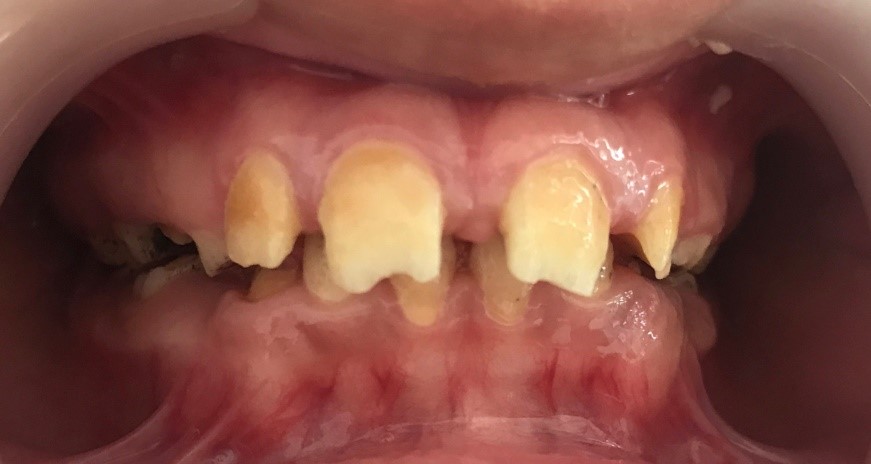
Figure 1: Pretreatment intraoral frontal view in occlusion showing generalized enamel defects affecting the mixed dentition, yellow-brown discoloration, post-eruptive enamel breakdown, severe anterior deep bite, and compromised dental esthetics.
Multiple retained primary second molars were present, while the permanent first molars exhibited severe enamel defects compromising both function and morphology. Two primary first molars (64,84) were considered non-restorable due to extensive structural damage (Figure 2).
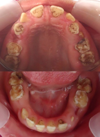
Figure 2: Pretreatment maxillary and mandibular occlusal views demonstrating generalized enamel hypoplasia with extensive occlusal breakdown, dentin exposure, and retained primary teeth
A renal ultrasonographic examination was also performed as part of the patient’s systemic assessment. The examination was within normal limits, with no visible renal calculi or signs of nephrocalcinosis. There was no dilatation of the pyelocaliceal cavities, proximal ureters, or ureters upstream of the bladder. The bladder had thin, regular walls and no abnormality was identified. These findings did not reveal any associated renal abnormality.
Radiographic examination, including panoramic and lateral cephalometric radiographs, confirmed the mixed dentition stage with generalized enamel defects affecting both the permanent and retained primary teeth.
The panoramic radiograph demonstrated reduced enamel radiopacity and extensive coronal involvement without evidence of associated developmental anomalies (Figure 3). The permanent successors of the extracted primary first molars were approaching eruption radiographically.
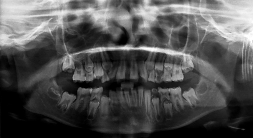
Figure 3: Pretreatment panoramic radiograph illustrating the mixed dentition stage, generalized enamel defects affecting both primary and permanent teeth and non-restorable primary first molars [teeth 64, and 74] presenting extensive structural destruction requiring extraction before definitive rehabilitation.
The lateral cephalometric radiograph was evaluated to assess the sagittal and vertical skeletal and dental relationships (Figure 4). Cephalometric analysis of the lateral radiograph showed an SNA angle of 81°, an SNB angle of 76°, an ANB angle of 5.0°, indicating a predominantly Class II skeletal relationship. The vertical skeletal pattern was essentially normodivergent (FMA, 26°). The IMPA was 96°, the FMIA was 76.5°, and the interincisal angle was 126°. Clinically, the patient presented with an Angle Class II Division 1 malocclusion, increased overjet, anterior deep overbite, and clinically evident proclination of the maxillary incisors. The apparent difference in overbite between the clinical frontal photograph and the lateral cephalometric image was interpreted in the context of differences in image acquisition and mandibular positioning.
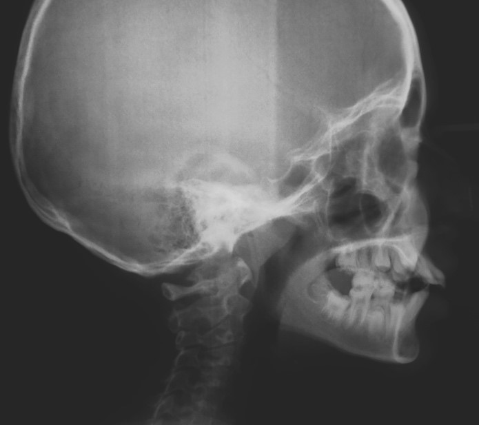
Figure 4: Pretreatment lateral cephalometric radiograph demonstrating a skeletal Class II relationship associated with a severe deep overbite, which motivated the initiation of interceptive orthodontic treatment following oral rehabilitation.
Oral hygiene was considered acceptable despite the patient’s difficulty in brushing because of dentin hypersensitivity. Periodontal examination showed healthy gingival tissues without signs of active periodontal disease.
Based on the clinical presentation, medical and dental history, and radiographic findings, a diagnosis of generalized non-syndromic amelogenesis imperfecta was established. The phenotype was characterized by generalized yellow-brown enamel defects, marked post-eruptive enamel breakdown, dentin exposure, and severe hypersensitivity, suggesting a predominantly hypocalcified phenotype. As no genetic or molecular testing was performed, the subtype was described according to its clinical phenotype rather than assigned to a definitive genetic diagnosis.
Treatment Objectives
The treatment objectives were to eliminate dentin hypersensitivity, preserve the remaining tooth structure, restore masticatory function, improve dental esthetics, re-establish the occlusal vertical dimension, and enhance the patient’s oral health-related quality of life.
Treatment Progress
Initial management consisted of oral hygiene reinforcement, individualized dietary counseling, professional prophylaxis, and impressions for diagnostic study casts.
The primary first molars 64 and 74 were considered non-restorable because of extensive structural damage and were extracted. No space maintainer was placed following extraction because radiographic examination showed that the corresponding permanent successors were approaching eruption. The expected duration requiring space maintenance was therefore considered limited. The eruption of the permanent successors was monitored clinically and radiographically during follow-up. After a two-week healing period, comprehensive oral rehabilitation was initiated.
Preformed stainless-steel crowns (3M™ ESPE™) were selected for the permanent first molars and retained primary second molars because of the extensive loss of enamel, the need for complete coronal protection, restoration of posterior occlusal support, and maintenance of the occlusal vertical dimension during growth.
Polycarbonate crowns (3M™ ESPE™) were selected for the permanent maxillary and mandibular incisors because they provided a conservative full-coverage option with satisfactory esthetics while requiring minimal removal of the remaining dental tissues.
The crowns were selected according to tooth size, adapted to the cervical margins, and evaluated for marginal adaptation, occlusion, and esthetic integration.
Both types of crowns were cemented using Meron glass ionomer luting cement (VOCO GmbH), according to the manufacturer’s recommendations. Excess cement was removed, and the occlusion was checked after cementation.
The posterior restorations provided complete coronal coverage, restored occlusal morphology, protected the remaining tooth structure from further wear, and re-established adequate posterior support and occlusal vertical dimension (Figure 5). Polycarbonate crowns were subsequently placed on the permanent maxillary and mandibular incisors to restore anterior tooth morphology and esthetics and to reduce dentin hypersensitivity (Figure 6).
Following restorative rehabilitation, interceptive orthodontic treatment was initiated using a Class II functional appliance (functional educator, Orthoplus) to address the developing malocclusion. The appliance was prescribed to reduce the anterior overjet and overbite and to improve tongue posture and the functional oral environment. It was introduced after stabilization of the dentition and restoration of posterior occlusal support.
The treatment sequence was designed to first stabilize and protect the compromised dentition, restore posterior occlusal support and the occlusal vertical dimension, and subsequently address the developing dental malocclusion through interceptive functional orthodontic therapy.
Written informed consent for the proposed treatment and publication of the clinical case and associated photographs was obtained from the patient’s legal guardian.
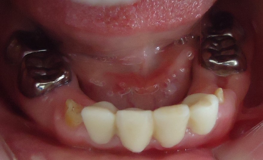
Figure 5: Placement of preformed stainless-steel crowns on the permanent first molars and retained primary second molars to restore posterior support, protect the remaining tooth structure, and re-establish the occlusal vertical dimension and placement of polycarbonate crowns on the permanent mandibular incisors to restore anterior esthetics and reduce dentin hypersensitivity
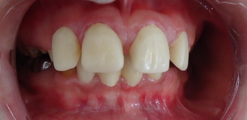
Figure 6: Post-treatment frontal intraoral view demonstrating improved dental esthetics, restoration of anterior tooth morphology, improved occlusal relationships, and successful comprehensive oral rehabilitation.
Treatment Results
At completion of the restorative phase, immediate improvement in dental esthetics, masticatory efficiency, and dentin hypersensitivity was observed. The patient and her parents reported high satisfaction with the treatment outcome. Clinical examination demonstrated satisfactory adaptation and integrity of the restorations, restoration of posterior occlusal support, and improvement of the anterior dental appearance. Oral hygiene remained satisfactory.
The patient was enrolled in a three-month recall program for clinical monitoring, oral hygiene reinforcement, topical fluoride application when indicated, evaluation of restoration integrity, and monitoring of the eruption of the permanent successors. At 18-month follow-up, the restorations remained clinically satisfactory, with maintained occlusal rehabilitation and no evidence of significant restoration failure.
Interceptive orthodontic follow-up was continued to monitor the development of the occlusion and the response to treatment. Definitive prosthetic rehabilitation was planned to be reconsidered after completion of craniofacial growth.
DISCUSSION
AI presents considerable restorative challenges because enamel defects compromise adhesion, accelerate tooth wear, and frequently result in dentin hypersensitivity, functional impairment, and poor esthetics from an early age [2,6]. Consequently, treatment planning should be individualized according to the patient’s age, clinical phenotype, severity of enamel defects, stage of dental development, and psychosocial needs. The primary objectives are to preserve tooth structure, relieve hypersensitivity, restore masticatory function, improve esthetics, and maintain the occlusal vertical dimension until definitive rehabilitation can be undertaken after completion of craniofacial growth [3,6].
The normal renal ultrasonographic findings in this patient are also noteworthy when considering the differential diagnosis of amelogenesis imperfecta. Although the clinical presentation was consistent with a non-syndromic form of AI, systemic assessment may be relevant when clinical findings raise the possibility of an associated syndrome. In the present case, renal ultrasonography showed no evidence of nephrocalcinosis, renal calculi, or urinary tract dilatation. This finding supports the absence of an identifiable renal abnormality in the systemic assessment performed in this patient. Nevertheless, the absence of renal abnormalities on ultrasonography should not be considered sufficient to establish a non-syndromic diagnosis when other clinical or genetic findings suggest a syndromic form.
The present case illustrates the importance of early intervention during the mixed dentition period. Delaying treatment may lead to progressive enamel loss, accelerated attrition, loss of vertical dimension, increased restorative complexity, and deterioration of oral health-related quality of life (OHRQoL). Recent evidence has emphasized that children with AI experience significantly lower OHRQoL than unaffected children because of pain, functional limitations, dissatisfaction with dental appearance, and reduced self-esteem [6,8,9]. In the present patient, hypersensitivity and the unaesthetic appearance of the dentition had negatively affected eating habits, oral hygiene practices, and social interactions, making comprehensive rehabilitation essential.
Several restorative options have been described for children with AI, including direct composite resin restorations, indirect adhesive restorations, polycarbonate crowns, strip crowns, zirconia crowns, stainless-steel crowns (SSCs), and, in selected cases, ceramic restorations [6,11]. However, during the mixed dentition stage, treatment should remain as conservative as possible because of the large pulp chambers, ongoing eruption, and continuous craniofacial growth. Therefore, interim restorations capable of protecting the remaining dental tissues while preserving future treatment options are generally recommended [3,11].
In the present case, SSCs were selected for the permanent first molars and retained primary second molars. These crowns remain the gold standard for restoring severely compromised posterior teeth in pediatric dentistry because they provide full coronal coverage, excellent durability, restoration of occlusal morphology, protection against further enamel breakdown, and maintenance of the occlusal vertical dimension [11,13,14]. Although esthetic posterior alternatives, such as prefabricated zirconia crowns, have recently gained popularity, SSCs continue to demonstrate superior longevity, lower technique sensitivity, and greater cost-effectiveness, particularly in children with extensive enamel defects [13,14].
Polycarbonate crowns were used to restore the permanent incisors because they provide a conservative, affordable, and esthetic interim solution during the mixed dentition stage. These restorations improve dental appearance, reduce dentin hypersensitivity, restore anterior function, and significantly enhance the patient’s self-confidence while preserving tooth structure for future definitive rehabilitation [3,11]. Similar favorable outcomes have been reported in previous clinical studies evaluating minimally invasive rehabilitation protocols for children with AI [7,11].
Re-establishing the occlusal vertical dimension constituted another important objective of treatment. Progressive enamel loss frequently leads to occlusal collapse, altered mastication, and increased functional overload. Full-coverage restorations on posterior teeth contribute to restoring occlusal stability while allowing physiological dentoalveolar development during growth [3,6]. In the present case, restoration of the vertical dimension resulted in improved masticatory efficiency and overall oral function without compromising future prosthetic management.
An additional consideration in this case was the premature loss of the primary first molars 64 and 74. Space maintenance was considered following extraction; however, a space maintainer was not placed because radiographic examination demonstrated that the corresponding permanent successors were approaching eruption. Under these circumstances, the anticipated period during which space maintenance would have been required was considered limited. The patient was therefore monitored clinically and radiographically for spontaneous eruption. This decision highlights the importance of individualized space-management planning after premature primary tooth loss. Although a lingual holding arch or other bilateral space-maintaining appliance may be appropriate in selected patients, the decision should take into account dental age, eruption stage, residual space, root development and the radiographic proximity of the permanent successors rather than being routinely applied after every premature extraction.
The orthodontic component of treatment also required careful interpretation. The patient presented clinically with an Angle Class II Division 1 dental relationship, increased overjet, anterior deep overbite, and clinically evident proclination of the maxillary incisors. Following restorative stabilization, an Orthoplus Class II functional educator was introduced as interceptive orthodontic treatment. The objectives were to reduce the increased overjet and overbite, improve the sagittal and vertical dental relationships, and encourage a more favorable tongue posture. The sequence of treatment was intentional: restoring posterior occlusal support and stabilizing the compromised dentition before functional orthodontic intervention provided a more stable dental environment for subsequent occlusal correction. This multidisciplinary sequencing is particularly relevant in children with AI, in whom orthodontic treatment must be integrated with the protection of structurally compromised teeth.
Beyond functional rehabilitation, early treatment also has an important psychosocial impact. Previous studies have demonstrated that children with AI frequently report embarrassment, social avoidance, reduced self-confidence, and emotional distress because of the appearance of their teeth [8-10].
Following rehabilitation, our patient reported complete resolution of hypersensitivity together with marked improvement in dental esthetics and self-esteem. These findings are consistent with previous reports showing significant improvements in oral health-related quality of life after comprehensive restorative treatment [9,10]. In addition to restorative rehabilitation, the patient was referred for interceptive orthodontic treatment to address the underlying skeletal Class II relationship and severe deep overbite. This multidisciplinary approach aimed not only to restore dental function and esthetics but also to optimize occlusal development during craniofacial growth.
Long-term follow-up remains essential because restorations placed during childhood should be regarded as transitional. Regular clinical and radiographic evaluations allow monitoring of eruption, occlusal development, restoration integrity, periodontal health, and oral hygiene. Definitive prosthetic rehabilitation should be considered only after completion of craniofacial growth, when more conservative adhesive ceramic or indirect restorations can be provided according to the patient’s functional and esthetic needs [3,6].
Overall, the present case supports current evidence advocating early multidisciplinary management of AI. Timely rehabilitation with minimally invasive full-coverage restorations effectively relieved hypersensitivity, restored function, improved esthetics, preserved tooth structure, and enhanced the patient’s quality of life while maintaining future restorative options.
CONCLUSION
Early comprehensive rehabilitation should be considered in children with amelogenesis imperfecta during the mixed dentition stage when enamel breakdown, hypersensitivity, functional impairment, or esthetic concerns compromise oral health and quality of life. In the present case, the combination of stainless-steel crowns for posterior teeth and polycarbonate crowns for anterior teeth provided conservative full-coverage rehabilitation, protected the remaining dental tissues, restored posterior occlusal support, reduced hypersensitivity, and improved dental esthetics and masticatory function.
Treatment planning should be individualized according to the severity of enamel defects, dental and skeletal development, eruption status, occlusal relationships, and psychosocial needs. Following premature extraction of primary teeth, the need for space maintenance should be assessed on an individual basis according to the proximity of the permanent successors and anticipated eruption time. Similarly, orthodontic diagnosis should distinguish dental from skeletal discrepancies and should be supported by appropriate clinical and cephalometric assessment when indicated.
Because these restorations are transitional, regular long-term clinical and radiographic follow-up is essential to monitor restoration integrity, eruption, occlusal development, oral hygiene, and the need for further restorative or orthodontic treatment. Longitudinal studies with standardized clinical, functional, occlusal, and quality-of-life outcomes are needed to evaluate the durability and effectiveness of conservative rehabilitation strategies for children with amelogenesis imperfect.
REFERENCES
- Sabandal MM, Schäfer E. (2016). Amelogenesis imperfecta: review of diagnostic findings and treatment concepts. Odontology. 104(3):245-256.
- Lundgren GP, Dahllöf G. (2024). Advances in clinical diagnosis and management of amelogenesis imperfecta in children and adolescents. J Dent. 147:105149.
- Dujic H, Bücher K, Schüler IM, Schmidt P, Hertel S, Timpel J, et al. (2025). Dental Management of Genetic Dental Disorders: A Critical Review. Journal of Dental Research. 104(4):369-379.
- Smith CEL, Poulter JA, Antanaviciute A, Kirkham J, Brookes SJ, Inglehearn CF, et al. (2017). Amelogenesis Imperfecta; Genes, Proteins, and Pathways. Frontiers in Physiology. 26;8:435.
- Witkop CJ. (1988). Amelogenesis imperfecta, dentinogenesis imperfecta and dentin dysplasia revisited: problems in classification. Journal of Oral Pathology. 17(9-10):547-553.
- Rhaiem M, Chalbi M, Bousaid S, Zouaoui W, Chemli MA. (2024). Dental treatment approaches of amelogenesis imperfecta in children and young adults: A systematic review of the literature. Journal of Esthetic and Restorative Dentistry. 36(6):881-891.
- McDonald S, Arkutu N, Malik K, Gadhia K, McKaig S. (2012). Managing the paediatric patient with amelogenesis imperfecta. British Dental Journal. 212(9):425-428.
- Parekh S, Almehateb M, Cunningham SJ. (2014). How do children with amelogenesis imperfecta feel about their teeth? International journal of Paediatric Dentistry. 24(5):326-335.
- Pousette Lundgren G, Karsten A, Dahllöf G. (2015). Oral health-related quality of life before and after crown therapy in young patients with amelogenesis imperfecta. Health and Quality of Life Outcomes. 13:197.
- Coffield KD, Phillips C, Brady M, Roberts MW, Strauss RP, Wright JT. (2005). The psychosocial impact of developmental dental defects in people with hereditary amelogenesis imperfecta. The Journal of the American Dental Association. 136(5):620-630.
- Marquezin MC, Zancopé BR, Pacheco LF, Gavião MB, Pascon FM. (2015). Aesthetic and functional rehabilitation of the primary dentition affected by amelogenesis imperfecta. Case Reports in Dentistry. 790890.
- Zagdwon AM, Fayle SA, Pollard MA. (2003). A prospective clinical trial comparing preformed metal crowns and cast restorations for defective first permanent molars. European journal of Paediatric Dentistry. 4(3):138-142.
- Innes NP, Ricketts D, Chong LY, Keightley AJ, Lamont T, Santamaria RM. (2015). Preformed crowns for decayed primary molar teeth. The Cochrane Database of Systematic Reviews. 2015(12):CD005512.
- Seale NS, Randall R. (2015). The use of stainless steel crowns: a systematic literature review. Pediatric Dentistry. 37(2):145-160.
