2026: Volume 8, Issue 1
Current Issue
 Abstract
  PDF
PDFOral Irritation Fibroma; Two Cases Report with Follow up and Literature Review
Alparslan Dilsiz1*, Suha Aksakal2, Ahmet Sanlıdag3, Abdullah Yavuz Ozmen4, Emrullah Yakut5
Received Date: August 09, 2026
Publication Date: August 24, 2026
Citation: Dilsiz A, et al. (2026). Oral Irritation Fibroma; Two Cases Report with Follow up and Literature Review. Dental. 8(1):25.
Copyright: Dilsiz A, et al. © (2026).
1Department of Periodontology, Faculty of Dentistry, Atatürk University, Erzurum/ TURKEY
2Department of Periodontology, Faculty of Dentistry, Atatürk University, Erzurum/ TURKEY
3Department of Periodontology, Faculty of Dentistry, Atatürk University, Erzurum/ TURKEY
4Department of Periodontology, Faculty of Dentistry, Atatürk University, Erzurum/TURKEY
5Department of Periodontology, Faculty of Dentistry, Atatürk University, Erzurum/ TURKEY
*Corresponding author: Alparslan DILSIZ, DDS, PhD. Professor, Department of Periodontology, Faculty of Dentistry, Atatürk University, 25240-Erzurum/ TURKEY, Fax: + 90 442 2361375 Tel: +90 533 4760078, E-mail: [email protected]; [email protected]; [email protected]
ABSTRACT
Aim: Irritation fibroma is a common benign reactive lesion of the oral mucosa that usually develops secondary to persistent mechanical irritation. Frequent etiological factors include chronic biting habits, foreign bodies, overhanging dental restorations, sharp tooth surfaces, and ill-fitting prostheses or other dental appliances. Habitual biting of the cheek, tongue, or lips represents a chronic, generally harmless form of self-inflicted trauma that may occur without the patient's awareness, often making the underlying cause difficult to identify during clinical examination.
The aim of the present study was to describe two cases of oral irritation fibroma and to present their clinical management together with the successful treatment outcomes.
Case Report: This case report presents the clinical characteristics, diagnostic evaluation, therapeutic management, and follow-up findings of two patients who presented with a gradually enlarging, painless lesion in the oral cavity. Both lesions were excised under local anesthesia, and the surgical specimens were submitted for histopathological analysis to establish the definitive diagnosis.
Results: The postoperative course was uneventful, with complete epithelialization achieved within 10 days. At subsequent follow-up examinations, both patients demonstrated satisfactory wound healing, and the surgical sites exhibited normal gingival architecture without any evidence of recurrence.
Conclusion: These cases emphasize the importance of comprehensive clinical evaluation, histopathological verification, and complete surgical excision in the successful management of irritation fibroma. Prompt diagnosis together with identification and elimination of the underlying etiological factors plays a key role in minimizing the risk of recurrence and achieving favorable clinical outcomes. Furthermore, patient adherence to treatment recommendations, appropriate psychological support when indicated, and regular dental follow-up visits may contribute to long-term disease control and maintenance of oral health.
Keywords: Irritation Fibroma, Traumatic Fibroma, Focal Fibrous Hyperplasia, Self-İnflicted, Biting, Chewing, Case Report
INTRODUCTION
Traumatic injuries of the oral cavity, including chemical, physical, and thermal insults, represent one of the most frequently encountered conditions affecting the oral soft tissues [1-3]. Irritation fibroma is a common benign reactive lesion that develops in response to persistent mechanical irritation and is also referred to as “traumatic fibroma” or “biting fibroma” [4,5]. Continuous local trauma caused by factors such as habitual cheek, lip, or tongue biting, sharp dental surfaces, overhanging restorations, foreign bodies, and ill-fitting prostheses or dental appliances may stimulate localized fibrous connective tissue proliferation.
Habitual biting of the cheek, tongue, or lips is generally considered a chronic, self-inflicted injury that is often harmless but may become clinically significant when persistent. Such behavior has frequently been associated with emotional disturbances or psychological stress [3-8]. The buccal mucosa is the most commonly affected site (40%), followed by the tongue (18%), lips (16.5%), and other locations, including the hard palate and gingiva (10%) [4-6]. Because the clinical appearance may resemble several other oral mucosal disorders, misdiagnosis is possible if the underlying habit is not recognized [9]. Moreover, many affected individuals are unaware of their parafunctional habit, which may complicate the diagnostic process.
Clinically, irritation fibroma usually presents as a solitary, smooth-surfaced, asymptomatic, painless, mucosa-colored lesion that may be sessile or pedunculated and can vary considerably in size [10]. In the literature, it has also been described using terms such as peripheral fibroma, fibrous nodule, fibroepithelial polyp, focal fibrous hyperplasia, and inflammatory fibrous hyperplasia, reflecting its mesenchymal connective tissue origin [4-9]. Reported prevalence ranges from 1% to 15%, and histologically the lesion consists of dense collagen bundles with mature fibroblasts without evidence of malignant transformation [11]. It is considered a localized, non-neoplastic reactive proliferation of fibrous connective tissue. Although irritation fibroma may occur at any age, including childhood and older adulthood, it has no established racial predilection and appears to be slightly more common among women over 30 years of age than men [4,5].
The development of irritation fibroma has been associated with a variety of chronic local irritants, including habitual biting, persistent inflammation, fixed or removable dental prostheses, foreign bodies, overhanging restorative margins, and sharp bony or dental prominences [11-15]. Despite its typically asymptomatic nature, the lesion may clinically resemble several reactive or neoplastic conditions, making careful differential diagnosis essential for appropriate management.
Treatment planning should be individualized according to the patient's age, lesion size, and clinical characteristics [4-9]. Some lesions, particularly those occurring during childhood, may regress spontaneously [4-6]. Small lesions that do not interfere with esthetics, speech, or mastication and are not subjected to repeated trauma or inflammation may be managed conservatively. However, symptomatic or persistent lesions generally require complete removal using techniques such as conventional surgical excision, electrosurgery, cryosurgery, or laser therapy [4-9]. Following complete excision and elimination of the underlying etiological factor, recurrence is uncommon [4].
The present study reports a case of biting fibroma and describes its clinical presentation, diagnosis, treatment, and successful postoperative outcome. This case report was prepared in accordance with the CaReL guidelines [16].
CASE REPORT
Case 1
A 36-year-old man was referred to the Department of Periodontology with complaints of pain and a gradually enlarging mass involving the left buccal mucosa.
During history taking, the patient acknowledged that he had been habitually chewing the inside of his left cheek for many years, although he had not previously recognized this behavior. He explained that the habit became more frequent during periods of psychological stress, with both the frequency and intensity of cheek biting increasing in parallel with his stress level. Apart from severe stress, there was no history of psychiatric illness. His medical history was otherwise unremarkable, and he was not receiving any regular medication. The lesion had enlarged slowly over several years and remained painless without tenderness.
Intraoral examination revealed a well-defined, pale pink, mobile soft-tissue mass located on the left buccal mucosa. The lesion measured approximately 20 mm in diameter and exhibited a smooth, non-ulcerated surface with a broad base and firm consistency. It was non-tender on palpation, and the overlying mucosa retained a normal color and texture (Figure 1A). No cervical lymphadenopathy or spontaneous bleeding was detected.
Panoramic radiographic examination demonstrated alveolar bone loss, dental caries, missing teeth, disruption of the lamina dura, widening of the periodontal ligament space, and furcation involvement (Figure 1B).
Periodontal examination revealed generalized gingival inflammation characterized by bleeding on probing, edema, and erythema associated with dental plaque accumulation. Additional findings included mild supragingival calculus deposits, increased probing depths, interproximal clinical attachment loss, tooth malposition, mobility, and the presence of carious lesions, while all teeth were non-tender to percussion.
Based on the clinical findings, a provisional diagnosis of biting fibroma was established.
Successful management required elimination of the contributing etiological factor, namely the patient's habitual cheek biting associated with psychological stress. Therefore, the patient was referred for psychiatric evaluation and counseling and was encouraged to discontinue the parafunctional habit.
Initial periodontal treatment included oral hygiene instruction, full-mouth scaling and root planing, followed by complete surgical excision of the lesion. The surgical wound was closed with 3-0 non-resorbable silk sutures (Figure 1C). The excised specimen (Figure 1D) was fixed in 10% formalin and submitted for histopathological examination. Microscopic evaluation demonstrated hyperplastic stratified squamous epithelium with hyperkeratosis and hyperchromatism of the basal and parabasal cell layers overlying dense fibrous connective tissue containing a mild chronic inflammatory infiltrate, confirming the diagnosis of irritation fibroma (Figure 1H).
Postoperatively, the patient was prescribed naproxen (550 mg every 12 hours for 5 days) for pain control and instructed to rinse twice daily with 0.12% chlorhexidine mouthwash (Kloroben®, Drogsan Drug Ltd., Istanbul, Turkey) for one week. He was also advised to avoid any mechanical trauma or excessive pressure at the surgical site during the healing period.
Complete soft-tissue healing was observed three weeks after surgery (Figure 1E). During approximately five years of postoperative follow-up, no evidence of recurrence was detected. At the follow-up visits, the patient reported that he had successfully discontinued his habitual cheek-chewing behavior, and the surgical outcome remained stable (Figure 1F and 1G).
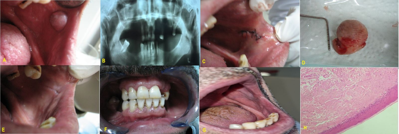
Figure 1: Case 1
A) Clinical aspect of the biting fibroma on the left check mucosa, B) Panoramic radiograph view, C) Clinical view of the surgical site, D) The view of excised tissue, E) Clinical view at 3 weeks post-surgery., F) Intraoral Clinical view at 5 years post-surgery., G) Clinical view at 5 years post-surgery, H) Histopathological appearance (H&E, ×200)
Case 2
A 65-year-old woman presented to the outpatient clinic with a painless mass located in the right maxillary anterior region that had been present for approximately 6–7 months (Figure 2A). Her medical history was unremarkable, and she was not receiving any regular medication. During the interview, the patient reported long-term use of a complete denture and stated that pain and swelling had developed after replacement with a new prosthesis. Clinical examination identified a well-defined, pedunculated nodular lesion measuring approximately 20 × 8 mm. The lesion exhibited a smooth surface and a color similar to the adjacent oral mucosa.
Based on the clinical examination, a provisional diagnosis of irritation fibroma was established, and complete excisional biopsy was planned (Figure 2B and 2C). The patient's prosthesis was also evaluated, followed by the necessary adjustment and polishing to eliminate potential sources of chronic irritation. Complete soft-tissue healing was observed 10 days after surgery (Figure 2D). During approximately 6 months of postoperative follow-up, no clinical evidence of recurrence was detected (Figure 2E). Histopathological examination demonstrated hyperplastic stratified squamous epithelium overlying dense collagenous connective tissue, confirming the diagnosis of irritation fibroma (Figure 2F).
The present case report was conducted in accordance with the ethical principles of the Declaration of Helsinki. Written informed consent was obtained from both patients for the publication of their clinical data and accompanying clinical photographs.
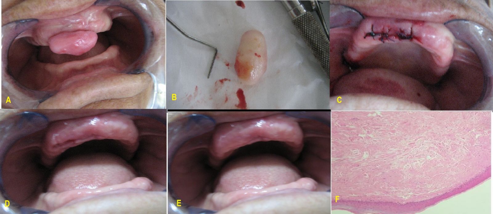
Figure 2: Case 2
A) Clinical aspect of the irritation fibroma, B) Clinical view of the surgical site, C) The view of excised tissue, D) Postoperative view after 10 days., E) Clinical view at 6 mounts post-surgery., F) Histopathological appearance (H&E, ×100)
DISCUSSION
Oral soft-tissue injuries may result from chemical, physical, or thermal trauma and represent an important group of non-plaque-induced gingival lesions. According to the 2017 World Workshop jointly organized by the European Federation of Periodontology (EFP) and the American Academy of Periodontology (AAP), traumatic lesions are classified among non-plaque-induced gingival diseases and may originate from physical, chemical, or thermal insults [1,2]. Self-inflicted injury constitutes one form of physical trauma and may arise from parafunctional habits such as biting, nibbling, rubbing, picking, scratching, sucking, or chewing of the oral soft tissues [6-12]. In the present case, chronic habitual cheek chewing was considered the principal etiological factor responsible for the development of the biting fibroma.
Habitual cheek or lip biting is generally regarded as an unconscious parafunctional behavior that is frequently associated with psychological conditions including stress, anxiety, and depression [10,11]. Previous studies have mainly described self-inflicted oral injuries in individuals with congenital syndromes, intellectual disabilities, or psychiatric disorders; however, similar lesions have also been reported in patients without these conditions, although such cases remain relatively uncommon [4,6,9]. In the present patient, severe psychological stress was considered the most likely trigger for the persistent cheek-chewing habit, which ultimately resulted in the development of the lesion.
Currently, no universally accepted treatment protocol exists for self-inflicted oral soft-tissue injuries. Management is often challenging because patient cooperation may be limited and discontinuation of the causative habit can be difficult to achieve [2,6,7]. These lesions frequently develop through a repetitive cycle in which chronic trauma induces inflammation, while the inflamed tissue becomes increasingly susceptible to further injury. Several authors have recommended the use of intraoral appliances, protective prostheses, behavioral modification, and, when necessary, sedation to minimize repetitive trauma and control destructive oral habits [4,6,8,9]. In the present case, psychiatric consultation was incorporated into the treatment plan, and the patient successfully discontinued the cheek-chewing habit following counseling.
The use of poorly adapted or ill-fitting dental prostheses has also been recognized as an important etiological factor contributing to the development of oral reactive lesions [7,8], as demonstrated in the second case presented in this report.
Clinically, irritation fibroma usually appears as a well-circumscribed, slow-growing, painless soft-tissue mass [4,6]. Although the lesion may occur bilaterally, its distribution often reflects the location of the underlying traumatic habit and may therefore be confined to a single site. Because its clinical appearance overlaps with several reactive and neoplastic lesions including peripheral giant cell granuloma, fibrous or hyperkeratotic polyps, salivary gland tumors, lipoma, mucocele, and neurofibroma histopathological examination is essential for establishing a definitive diagnosis [4,6]. In the present report, the biting fibroma was located unilaterally on the left buccal mucosa and exhibited the characteristic clinical features of a well-defined, painless lesion.
Complete surgical excision was selected as the treatment of choice for both patients. Several therapeutic modalities have been described for the management of irritation fibroma, including conventional surgical excision, electrosurgery, cryosurgery, and laser-assisted surgery [9-12]. In the present cases, conventional surgical excision resulted in satisfactory healing without postoperative complications, and no recurrence was detected throughout the follow-up period, supporting previous reports that complete removal of the lesion together with elimination of the underlying etiological factor provides a favorable long-term prognosis.
CONCLUSIONS
Although oral traumatic lesions associated with habitual cheek, lip, and tongue biting are generally considered relatively uncommon, they represent an important clinical entity that should not be overlooked in routine dental practice. The present cases underscore the importance of comprehensive clinical evaluation, histopathological confirmation, and complete surgical excision for the successful management of irritation fibroma. Prompt diagnosis together with identification and elimination of the underlying etiological factors is essential to minimize the risk of recurrence and achieve favorable long-term clinical outcomes. Furthermore, patient adherence to treatment recommendations, appropriate psychological support when indicated, and regular dental follow-up examinations may contribute substantially to long-term disease control and maintenance of oral health.
ACKNOWLEDGEMENTS
Authors’ Contributions
Alparslan Dilsiz diagnosed and managed the patients, conceived the idea of the case report, drafted the manuscript, and critically revised the manuscript
Suha Aksakal, Ahmet Sanlıdag, Abdullah Yavuz Ozmen, and Emrullah Yakut performed periodontal therapy and surgery procedure. All authors read and approved final version of text.
Funding
This study received no specific grant from any funding agency in the public, commercial, or non-profit sectors.
Competing Interests
The authors declare no competing interests.
REFERENCES
- Holmstrup P, Plemons J, Meyle J. (2018). Non-plaque-induced gingival diseases. J Periodontol. 89 Suppl 1:S28-S45.
- Dilsiz A, Sevinç Gü SNL. (2023). Investigation of Biopsied Non-Plaque-Induced Gingival Lesions in a Turkish Population: A 5-Year Retrospective Study. The Eurasian Journal of Medicine. 55(2):100-103.
- Dilsiz A, Aydin T. (2009). Self-inflicted gingival injury due to habitual fingernail scratching: a case report with a 1-year follow up. European Journal of Dentistry. 3(2):150-154.
- Srivastava A, Bhati P, Khanna V, Arora K, Saxena R. (2024). A Large Irritation Fibroma of the Hard Palate: A Rare Clinical Entity. Cureus. 16(6):e62244.
- Diwan B, Shirbhate U, Bajaj P, Reche A, Pahade A. (2023). Conventional Scalpel and Diode Laser Approach for the Management of Traumatic Fibroma. Cureus. 15(10):e47810.
- Cohen PR. (2022). Biting Fibroma of the Lower Lip: A Case Report and Literature Review on an Irritation Fibroma Occurring at the Traumatic Site of a Tooth Bite. Cureus. 14(12):e32237.
- de Santana Santos T, Martins-Filho PR, Piva MR, de Souza Andrade ES. (2014). Focal fibrous hyperplasia: A review of 193 cases. Journal of Oral and Maxillofacial Pathology. 18(Suppl 1):S86-S89.
- Mujica V, Rivera H, Carrero M. (2008). Prevalence of oral soft tissue lesions in an elderly venezuelan population. Medicina oral, patología oral y cirugía bucal. 13(5):E270-E274.
- Limeres J, Feijoo JF, Baluja F, Seoane JM, Diniz M, Diz P. (2013). Oral self-injury: an update. Dental Traumatology. 29(1):8-14.
- Fatima R, Abid K, Baig NN, Ahsan SB. (2019). Association of cheek-biting and depression. Journal of the Pakistan Medical Association. 69(1):49-52.
- Bhatia NK. (2015). Morsicatiobuccarum and labiorum with depression. Int J Dent Health Sci. 2:1639-1642.
- Bakhtiari S, Taheri JB, Sehhatpour M, Asnaashari M, Attarbashi Moghadam S. (2015). Removal of an Extra-large Irritation Fibroma With a Combination of Diode Laser and Scalpel. J Lasers Med Sci. 6(4):182-184.
- Bhatia SK, Goyal A, Kapur A. (2013). Habitual biting of oral mucosa: A conservative treatment approach. Contemporary Clinical Dentistry. 4(3):386-389.
- Romero M, Vicente A, Bravo LA. (2005). Prevention of habitual cheek biting: a case report. Spec Care Dentist. 25(4):214-216.
- Walker RS, Rogers WA. (1992). Modified maxillary occlusal splint for prevention of cheek biting: a clinical report. The Journal of Prosthetic Dentistry. 67(5):581-582.
- Prasad S, Nassar M, Azzam AY, Jose FG, Jamee M, Sliman RKA.et al. (2024). CaReL Guidelines: A Consensus-Based Guidelines on Case Reports and Literature Review (CaReL). Barw Medical Journal. 2(2):13-19.
