2026: Volume 8, Issue 1
Current Issue
 Abstract
  PDF
PDFManagement of Two Erupted Mesiodens Associated with Maxillary Central Incisor Malposition: A Case Report
Farah Chouchene1,2*, Nehed Sboui3
1Associate Professor, Department of Dentistry, Ibn El Jazzar University Hospital, Kairouan, Faculty of Dental Medicine of Monastir. Tunisia
2Laboratory of Biological, Clinical and Dento-Facial Approach, University of Monastir, Monastir, Tunisia
3Specialist in Pediatric Dentistry. University of Monastir, Monastir, Tunisia
*Correspondent author: Farah Chouchene, Associate Professor, Department of Dentistry, Ibn El Jazzar University Hospital, Kairouan, Faculty of Dental Medicine of Monastir. Tunisia, Email: [email protected]
Received Date: September 02, 2026 Publication Date: September 22, 2026
Citation: Chouchene F, Sboui N. (2026). Management of Two Erupted Mesiodens Associated with Maxillary Central Incisor Malposition: A Case Report. Dental. 8(1):27.
Copyright: Chouchene F and Sboui N, et al. © (2026).
1Associate Professor, Department of Dentistry, Ibn El Jazzar University Hospital, Kairouan, Faculty of Dental Medicine of Monastir. Tunisia
2Laboratory of Biological, Clinical and Dento-Facial Approach, University of Monastir, Monastir, Tunisia
3Specialist in Pediatric Dentistry. University of Monastir, Monastir, Tunisia
*Correspondent author: Farah Chouchene, Associate Professor, Department of Dentistry, Ibn El Jazzar University Hospital, Kairouan, Faculty of Dental Medicine of Monastir. Tunisia, Email: [email protected]
ABSTRACT
Mesiodens are supernumerary teeth that may interfere with the eruption, position, and alignment of permanent maxillary incisors. The occurrence of two erupted mesiodens associated with established displacement and rotation of the permanent central incisors presents a particular diagnostic and therapeutic challenge. An 11-year-old girl presented with two erupted, palatally positioned mesiodens associated with ectopic positioning, axial displacement, and rotation of the permanent maxillary central incisors. One mesiodens was conical and the other showed mild dilaceration. Periapical radiography confirmed their position and relationship with the adjacent incisors. Both mesiodens were surgically removed under local anesthesia. An approximately 8-mm maxillary midline diastema with persistent central incisor malposition and rotation remained after extraction. Sectional fixed orthodontic treatment was initiated two weeks later. Following a change in the axial inclination of the central incisors during treatment, the orthodontic mechanics were modified and a transpalatal arch was introduced to reinforce posterior anchorage. After 12 months of active orthodontic treatment, complete closure of the diastema and satisfactory alignment of the maxillary central incisors were achieved. This case highlights that removal of erupted double mesiodens may not be sufficient when significant positional abnormalities of the permanent incisors have already developed. Sequential surgical removal followed by individualized orthodontic treatment and appropriate anchorage control can provide satisfactory correction while avoiding surgical exposure and traction of the permanent incisors.
Keywords: Mesiodens, Supernumerary Teeth, Double Mesiodens, Maxillary Central Incisors, Orthodontic Treatment, Dental Malalignment, Case Report
INTRODUCTION
Mesiodens are developmental dental anomalies characterized by the presence of supernumerary teeth in the maxillary anterior region, most commonly between the permanent central incisors. They represent the most common type of supernumerary tooth and may remain asymptomatic or cause complications including delayed or ectopic eruption, displacement, rotation, crowding, diastema, and, in some cases, root resorption of adjacent permanent teeth [1-3].
The clinical presentation of mesiodens is variable, ranging from an incidental radiographic finding to a fully erupted tooth associated with anterior malocclusion or occlusal interference [1-3]. The presence of two mesiodens, often referred to as double mesiodens, is considerably less common than the occurrence of a single mesiodens and may be associated with disturbances in eruption and anterior tooth position [4,5].
The etiology of mesiodens remains incompletely understood. Proposed mechanisms include localized hyperactivity of the dental lamina, dichotomy of the tooth germ, and multifactorial genetic and environmental influences [1,4]. Recent genetic studies have identified associations between mesiodens and variants in genes including FREM2 and PTPN23; however, these findings do not establish a specific genetic etiology in individual patients [6,7].
Accurate diagnosis and appropriate radiographic assessment are important for determining the position, morphology, and relationship of mesiodens to adjacent permanent teeth. Conventional periapical and panoramic radiographs can provide essential diagnostic information, whereas cone-beam computed tomography (CBCT) may be considered when three-dimensional imaging is expected to influence diagnosis or treatment planning [5,8].
Management depends on the patient’s age, tooth position, morphology, eruption status, and the presence and severity of associated dental disturbances. Treatment options range from clinical and radiographic observation in selected cases to surgical removal, with orthodontic treatment when displacement, rotation, space discrepancy, or other malocclusion persists [1,3,9,10]. In erupted mesiodens, surgical removal may directly eliminate the local mechanical obstruction; however, when permanent incisors have already developed significant displacement or rotation, orthodontic intervention may subsequently be required to restore alignment and space relationships [3,9,10].
Although double mesiodens have been described, published reports have frequently focused on impacted or unerupted supernumerary teeth and on their effects on the eruption of permanent incisors [4,9-13]. The present case is clinically distinctive because it involves two simultaneously erupted, palatally positioned mesiodens in an otherwise nonsyndromic child, associated with established ectopic positioning, axial displacement, and rotation of both permanent maxillary central incisors. In addition, the permanent incisors were already erupted and therefore did not require surgical exposure or orthodontic traction. The case illustrates the clinical decision-making involved in combining surgical removal with subsequent orthodontic correction and adapting anchorage control according to the orthodontic response.
This case report describes the sequential surgical and orthodontic management of an 11-year-old girl with two erupted, palatally positioned mesiodens associated with established ectopic positioning, axial displacement, and rotation of the permanent maxillary central incisors.
CASE PRESENTATION
This case report was prepared in accordance with the CARE (CAse REport) Guidelines [11].
Written informed consent was obtained from the patient’s parents for publication of the case report and accompanying clinical photographs and radiographs.
The timeline of clinical interventions is summarized in Table 1.
Table 1: Timeline of clinical management and follow-up
|
Time point |
Clinical event |
|---|---|
|
Initial Presentation |
Diagnosis of two erupted palatally positioned mesiodens and malposition of the permanent maxillary central incisors |
|
Day 0 |
Surgical extraction of both mesiodens under local anesthesia |
|
Two weeks after extraction |
Initiation of sectional fixed orthodontic treatment |
|
Four months after initiation of orthodontic treatment |
Re-evaluation of tooth axial inclination; modification of orthodontic mechanics and placement of a transpalatal arch for anchorage reinforcement. |
|
12 months after initiation of orthodontic treatment |
Complete closure of the 8 mm maxillary midline diastema and satisfactory alignment of the central incisors |
Diagnosis and Etiology
An 11-year-old girl was referred to the Department of Pediatric and Preventive Dentistry at the Faculty of Dental Medicine of Monastir for evaluation of two supernumerary teeth in the maxillary anterior region. The patient’s primary concern was the esthetic appearance of her smile. She reported being teased at school because of the appearance of her anterior teeth.
Her medical history was unremarkable, and no relevant family history of supernumerary teeth was reported. There was no history of previous dental trauma or clinical evidence suggestive of an associated syndrome. Parental consanguinity was reported. The patient did not report spontaneous pain but described discomfort during biting.
Extraoral examination revealed a convex facial profile, competent lips, an acute nasolabial angle, and hyperactivity of the mentalis muscle.
Intraoral examination during the mixed dentition stage revealed two erupted supernumerary teeth located palatally to the maxillary permanent central incisors. The permanent maxillary central incisors exhibited marked ectopic positioning, axial displacement, and rotation. One supernumerary tooth was conical, whereas the other exhibited mild dilaceration (Figure 1).
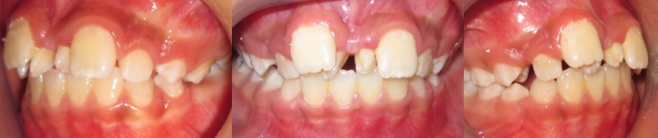
Figure 1: Intraoral photographs showing the two erupted supernumerary teeth positioned palatally between the maxillary central incisors and the associated malalignment of the maxillary anterior teeth.
An anterior periapical radiograph confirmed the presence of two supernumerary teeth and demonstrated their relationship with the adjacent permanent central incisors. Both supernumerary teeth had shorter roots than the adjacent central incisors (Figure 2).
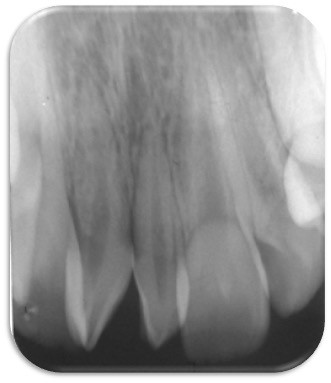
Figure 2: Anterior periapical radiographs demonstrating the presence and position of the two mesiodens between the maxillary central incisors.
Because both teeth were clinically erupted and their position and relationship with the adjacent incisors could be adequately assessed using conventional radiography, CBCT was not performed.
Based on the clinical and radiographic findings, the diagnosis was two erupted, palatally positioned mesiodens associated with ectopic positioning, axial displacement, and rotation of the permanent maxillary central incisors.
The etiology of the mesiodens could not be established definitively. There was no history of dental trauma, no evidence of an associated syndrome, and no family history of supernumerary teeth. Although parental consanguinity was reported, no genetic investigation was performed; therefore, no specific genetic contribution could be inferred.
Treatment Objectives
The treatment objectives were to eliminate the mechanical interference caused by the two erupted mesiodens, prevent further displacement of the permanent maxillary central incisors, and restore satisfactory anterior dental alignment. Because the central incisors were already ectopically positioned, axially displaced, and rotated, removal of the mesiodens alone was considered unlikely to provide complete correction. A sequential surgical and orthodontic approach was therefore planned.
The surgical phase aimed to remove the supernumerary teeth and eliminate the local obstruction. The subsequent orthodontic phase aimed to close the residual maxillary midline diastema, correct the position, axial inclination, and rotation of the central incisors, and establish satisfactory anterior alignment and occlusal function. Anchorage control was incorporated into the treatment plan to minimize unwanted posterior tooth movement during anterior space closure. Esthetic improvement of the anterior dentition was also considered an important treatment objective.
Treatment Progress
Because both mesiodens were erupted, palatally positioned, and in close proximity to the permanent maxillary central incisors, surgical removal was selected as the initial treatment phase. This approach was preferred to observation because the supernumerary teeth were already causing clinically significant interference and were associated with established displacement and rotation of the permanent incisors. Both mesiodens were extracted under local anesthesia to eliminate the obstruction and facilitate subsequent orthodontic correction (Figure 3A and B).
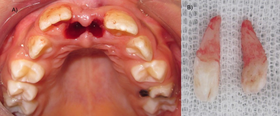
Figure 3: Extraction of the mesiodens. (A) Intraoral photograph showing the extraction sites following surgical removal of the two mesiodens. (B) Photograph of the two extracted mesiodens.
Following extraction, an approximately 8-mm maxillary midline diastema remained between the permanent central incisors, which continued to exhibit ectopic positioning, axial displacement, and rotation. As these positional abnormalities persisted after removal of the mechanical obstruction, orthodontic treatment was considered necessary rather than relying on spontaneous correction.
Orthodontic treatment was initiated two weeks after surgical removal using a sectional fixed orthodontic appliance (Figure 4A).
Metal brackets were bonded to the maxillary permanent central incisors (11 and 21) and the first premolars (14 and 24), as the remaining permanent teeth were still erupting. This sectional approach allowed focused correction of the affected anterior teeth while limiting appliance involvement in the developing dentition. Transparent orthodontic elastics were used for ligation and to assist with anterior tooth alignment.
A 0.012-inch round nickel-titanium (NiTi) archwire was initially placed to provide light, continuous forces for progressive alignment. The archwire was subsequently replaced at approximately monthly appointments, progressing from a 0.012-inch to a 0.014-inch and then a 0.016-inch round NiTi archwire. The patient was reviewed monthly for clinical assessment and orthodontic activation (Figure 4B).
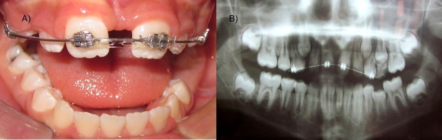
Figure 4: Progression of orthodontic treatment. (A) Intraoral photograph showing progressive closure of the maxillary midline diastema. (B) Panoramic radiograph demonstrating progressive space closure and an alteration in long axis of the maxillary central incisors during orthodontic treatment.
After four months of orthodontic treatment, a change in the long axis of the maxillary central incisors was observed. The orthodontic mechanics were therefore modified while maintaining the brackets on the central incisors (Figure 5A). A transpalatal arch was subsequently placed to stabilize the first permanent molars and reinforce posterior anchorage. This modification was intended to minimize unwanted posterior tooth movement and preserve the remaining space required for continued anterior correction and diastema closure (Figure 5B).
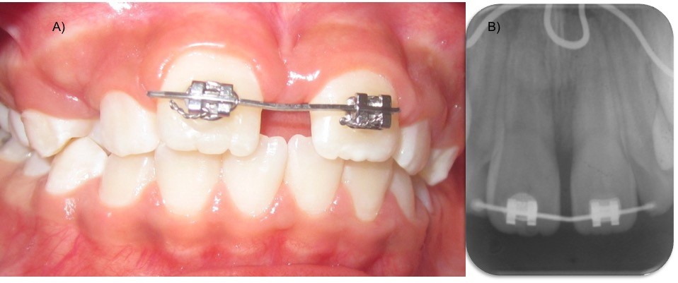
Figure 5: Modification of the orthodontic mechanics and anchorage reinforcement. (A) Intraoral photograph showing the modified orthodontic mechanics following alteration of the long axis of the maxillary central incisors. (B) Periapical radiograph showing the brackets bonded to the maxillary central incisors and the transpalatal arch used to reinforce posterior. anchorage and prevent unwanted space loss during orthodontic correction.
Orthodontic treatment was continued with progressive control of the maxillary central incisors and closure of the residual anterior space. During the finishing phase, a rectangular 0.016 × 0.022-inch stainless-steel archwire was used to improve three-dimensional control of tooth position and axial inclination.
After a total of 12 months of active orthodontic treatment, the desired clinical correction was achieved and the fixed appliance was removed (Figure 6A and B).
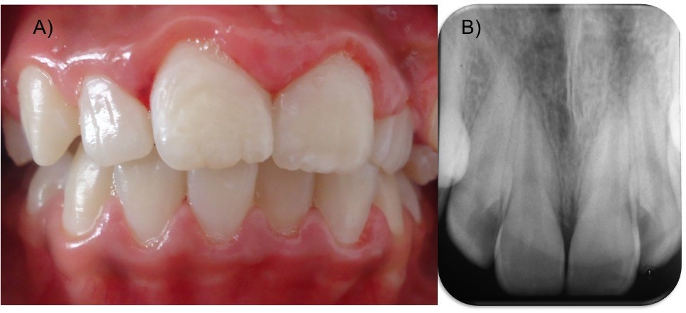
Figure 6: Final clinical and radiographic outcomes after 12 months of orthodontic treatment. (A) Final intraoral photograph showing complete closure of the maxillary midline diastema and satisfactory alignment of the maxillary central incisors. (B) Periapical radiograph showing the final position and root alignment of the maxillary central incisors.
Treatment Outcome
The combined surgical and orthodontic treatment resulted in a satisfactory clinical outcome. At completion of the 12-month orthodontic treatment, the approximately 8-mm maxillary midline diastema was completely closed, with satisfactory alignment of the permanent maxillary central incisors. The treatment corrected not only the residual space discrepancy but also the pre-existing ectopic positioning, axial displacement, and rotation of the central incisors.
A panoramic radiograph obtained during treatment demonstrated progressive orthodontic space closure and correction of the position of the maxillary central incisors. A subsequent periapical radiograph demonstrated the orthodontic brackets on the maxillary central incisors and the transpalatal arch used for posterior anchorage control. Final intraoral photographs confirmed complete closure of the anterior diastema and satisfactory alignment of the maxillary central incisors after completion of active treatment.
The patient reported satisfaction with the improvement in the appearance of her smile following treatment. No adverse events or complications were reported during the surgical or orthodontic treatment period.
DISCUSSION
The present case illustrates an uncommon clinical presentation of two erupted mesiodens associated with established positional abnormalities of the permanent maxillary central incisors. Although mesiodens are the most common type of supernumerary tooth, the occurrence of two mesiodens in the maxillary anterior region is considerably less frequent and may result in disturbances of eruption, tooth position, and anterior occlusion [4,5]. The present case is distinctive because both supernumerary teeth were erupted and palatally positioned, while the permanent maxillary central incisors had already developed marked ectopic positioning, axial displacement, and rotation.
The clinical consequences observed in this patient are consistent with the recognized effects of mesiodens on the developing permanent dentition. Supernumerary teeth may interfere with the normal eruption pathway and may be associated with delayed or ectopic eruption, displacement, rotation, crowding, diastema, and other positional abnormalities of adjacent permanent teeth [1-4].
In the present case, the simultaneous presence of two erupted mesiodens within the limited space of the premaxillary region was associated with substantial disruption of the position and alignment of both permanent central incisors.
The morphology and anatomical position of the mesiodens were also clinically relevant. One supernumerary tooth was conical, whereas the other exhibited mild dilaceration. Supernumerary teeth demonstrate considerable morphological and positional variability, and their morphology, orientation, and location may influence their clinical presentation and management [5,8]. The palatal position observed in the present patient is consistent with the range of anatomical presentations reported for mesiodens [5,8]. Because both supernumerary teeth were erupted, they could be directly identified clinically and removed without the need for surgical exposure of an unerupted supernumerary tooth.
The presence of multiple supernumerary teeth also emphasizes the importance of evaluating the entire anterior dentition when a supernumerary tooth is identified. In a radiographic study of a Saudi population, 24% of affected patients presented with two supernumerary teeth [12]. This finding should not be interpreted as the prevalence of double mesiodens because the study evaluated supernumerary teeth more broadly. Nevertheless, it supports the clinical importance of carefully assessing the surrounding dentition for additional supernumerary teeth when one is identified [12].
An important feature of this case was the persistence of significant anterior malposition after removal of the mesiodens. An approximately 8-mm maxillary midline diastema remained, and the permanent central incisors continued to demonstrate ectopic positioning, axial displacement, and rotation. This finding emphasizes the distinction between removing the local mechanical obstruction and correcting the orthodontic consequences that have already developed. Although spontaneous improvement in the position of permanent incisors may occur after removal of a supernumerary tooth in selected patients, orthodontic intervention may be required when significant displacement, rotation, or space discrepancy persists [3,9]. Evidence from a systematic review and meta-analysis of interventions for impacted maxillary incisors associated with supernumerary teeth also supports additional orthodontic intervention when spontaneous eruption or satisfactory alignment does not occur [13].
The timing of mesiodens removal should be individualized according to the patient’s age, eruption status, morphology and position of the supernumerary tooth, stage of development of the adjacent permanent teeth, and associated complications [9,10]. Current evidence does not support a single timing protocol applicable to every patient, and the potential benefits of intervention must be balanced against the risks of surgical intervention near developing permanent teeth [10,14]. In the present case, both mesiodens were already erupted and were directly interfering with the permanent central incisors. Their removal was therefore undertaken because they represented an established mechanical obstruction associated with clinically significant tooth displacement rather than solely because they were supernumerary teeth [10,14].
The classification and description of supernumerary teeth may also have clinical relevance. Recent literature has emphasized considerable variation in the terminology and classification of supernumerary teeth and has proposed a revised framework incorporating their morphological and positional characteristics [15]. In the present case, the two supernumerary teeth displayed different morphological features, with one being conical and the other exhibiting mild dilaceration. Accurate description of these characteristics is relevant because morphology and position may influence both the clinical consequences of supernumerary teeth and their management [5,8,15].
The etiological significance of the reported parental consanguinity requires cautious interpretation. The etiology of mesiodens remains incompletely understood and is considered multifactorial, with developmental and genetic mechanisms potentially contributing to their formation [1,4]. Recent genetic studies have identified associations between mesiodens and variants in FREM2 and PTPN23, supporting a possible genetic contribution to supernumerary tooth development [6,7]. Familial nonsyndromic occurrence of mesiodens has also been documented, including families in which multiple affected individuals presented without an associated syndrome [16]. Nevertheless, familial occurrence and genetic associations identified in research cohorts cannot establish causality in an individual patient. In the present case, no genetic investigation was performed, and neither a relevant family history nor syndromic features were identified. Accordingly, parental consanguinity should be regarded as a clinical characteristic rather than evidence of a specific hereditary mechanism [6,7,16].
Radiographic assessment was essential for confirming the diagnosis and evaluating the relationship between the supernumerary teeth and the permanent central incisors. In the present patient, periapical radiography provided sufficient information because both mesiodens were clinically erupted and their position and relationship with the adjacent teeth could be adequately assessed using conventional imaging. Contemporary radiological approaches emphasize selecting the imaging modality according to the diagnostic question, with three-dimensional imaging reserved for situations in which additional information is expected to influence diagnosis, surgical access, or treatment planning [5,8]. Avoiding CBCT in this patient was therefore based on the adequacy of conventional radiographic information rather than on a general contraindication to CBCT in children [8].
The orthodontic treatment in the present case was planned as a complementary second phase rather than as an alternative to surgical removal. A sectional fixed appliance was selected because the treatment objectives extended beyond simple space closure and required controlled correction of the position and axial inclination of the permanent central incisors. Light initial nickel-titanium archwires were used for progressive alignment, followed by a rectangular stainless-steel archwire to provide greater three-dimensional control during the finishing phase. This approach was consistent with the need to address established positional abnormalities after removal of the obstructing supernumerary teeth [3,9,13].
The introduction of a transpalatal arch after four months of orthodontic treatment represents an important biomechanical adaptation in this case. A change in the long axis of the maxillary central incisors was observed during treatment, prompting modification of the orthodontic mechanics. The transpalatal arch was subsequently used to stabilize the first permanent molars and reinforce posterior anchorage while anterior correction continued. This sequence illustrates the importance of monitoring orthodontic response and adapting treatment mechanics according to the observed tooth movement. Anchorage control is particularly relevant when anterior space closure is required and unwanted posterior tooth movement could compromise the intended correction [3,9].
The final outcome was favorable, with complete closure of the approximately 8-mm maxillary midline diastema and satisfactory alignment of the permanent central incisors after 12 months of active orthodontic treatment. The correction involved not only space closure but also improvement in the pre-existing ectopic positioning, axial displacement, and rotation of the central incisors.
Previous reports have described surgical removal of mesiodens followed by orthodontic management when persistent eruption or alignment disturbances remain [3,4,9,13,14]. The present case differs from many reports involving impacted permanent incisors because both mesiodens were erupted, the permanent central incisors were already present and displaced, and treatment did not require surgical exposure or orthodontic traction of an impacted central incisor [4,9,13].
The psychosocial dimension of the case is also clinically relevant. The patient initially sought treatment because of concern about the appearance of her smile and reported being teased at school because of her anterior dental appearance. Visible developmental dental anomalies and anterior malocclusion may affect children’s psychosocial well-being, including perceptions of dental appearance and social interactions [1,3]. Although no validated patient-reported outcome instrument was used in this case, the patient reported satisfaction with the improvement in the appearance of her smile following treatment. This observation should therefore be interpreted as a patient-reported clinical experience rather than as a formally measured improvement in psychological well-being.
Several limitations should be acknowledged. First, this is a single case report, and the outcome cannot establish the superiority of a particular surgical or orthodontic protocol or be generalized to all patients with double mesiodens. Second, although parental consanguinity was documented, no genetic investigation was performed; therefore, no specific hereditary mechanism can be inferred. Third, no validated patient-reported outcome instrument was used to assess the psychosocial impact of treatment. Finally, longer-term follow-up would be desirable to assess the stability of the orthodontic correction and the periodontal and root status of the permanent maxillary central incisors.
Overall, this case demonstrates that the management of erupted double mesiodens may require more than surgical removal when substantial positional abnormalities of the permanent central incisors have already developed. In such circumstances, treatment should address both the local mechanical obstruction and its established orthodontic consequences. In the present patient, sequential surgical removal followed by sectional fixed orthodontic treatment, with subsequent reinforcement of posterior anchorage using a transpalatal arch, resulted in complete closure of the anterior diastema and satisfactory alignment of the maxillary central incisors within 12 months.
CONCLUSION
Double mesiodens can significantly interfere with the development and alignment of the maxillary anterior dentition. Surgical removal of erupted mesiodens eliminates local obstruction but may not spontaneously resolve established displacement and rotation of central incisors. Sequential sectional fixed orthodontic treatment, combined with appropriate anchorage control using a transpalatal arch, provides an effective approach for managing complex space closure and alignment within 12 months.
REFERENCES
- Russell KA, Folwarczna MA. (2003). Mesiodens—diagnosis and management of a common supernumerary tooth. Journal - Canadian Dental Association. 69(6):362-366.
- Asaumi JI, Shibata Y, Yanagi Y, Hisatomi M, Matsuzaki H, Konouchi H, et al. (2004). Radiographic examination of mesiodens and their associated complications. Dento Maxillo Facial Radiology. 33(2):125-127.
- Ayers E, Kennedy D, Wiebe C. (2014). Clinical recommendations for management of mesiodens and unerupted permanent maxillary central incisors. European Archives of Paediatric Dentistry. 15(6):421-428.
- Bhatara S, Goswami M, Shivangani, Rahman B, Gogoi A. (2024). Double mesiodens in the mixed dentition of non-syndromic North-Indian patients: a case series. Cureus. 16(2):e54161.
- Lo-Casto A, Panzarella V, Palizzolo E, et al. (2025). Radiological features of double and inverted mesiodens: a systematic review of the literature and presentation of two cases. Medicina oral, patología oral y cirugía bucal. 30(6):e906-e915.
- Kantaputra PN, Tripuwabhrut K, Anthonappa RP, Chintakanon K, Ngamphiw C, Adisornkanj P, et al. (2023). Heterozygous variants in FREM2 are associated with mesiodens, supernumerary teeth, oral exostoses, and odontomas. Diagnostics (Basel). 13(7):1214.
- Adisornkanj P, Chanprasit R, Eliason S, Fons JM, Intachai W, Tongsima S, et al. (2023). Genetic variants in protein tyrosine phosphatase non-receptor type 23 are responsible for mesiodens formation. Biology (Basel). 12(3):393.
- Mallineni SK, Anthonappa RP, Jayaraman J, King NM. (2025). Radiographic localization of supernumerary teeth: a narrative review. Frontiers in Dental Medicine. 6:1495025.
- Shetty V, Shetty RY, Mehrotra D, Usman A, Prabhu N. (2024). Management of impacted and inverted mesiodens: a report of two cases. Journal of Pharmacy and Bioallied Sciences. 16:S4120-S4122.
- Adachi M. Mesiodens removal: resolving the early versus late intervention dilemma. (2026). J Oral Maxillofac Surg. 84(2):225-232.
- Riley DS, Barber MS, Kienle GS, Aronson JK, von Schoen-Angerer T, Tugwell P, et al. (2017). CARE guidelines for case reports: explanation and elaboration document. J Clin Epidemiol. 89:218-235.
- Mallineni SK, Aldhuwayhi S, Deeban Y, Almutairi KS, Alhabrdi SN, Almidaj MA, et al. (2024). Prevalence, occurrence, and characteristics of supernumerary teeth among the Saudi Arabian population using panoramic radiographs. Diagnostics (Basel). 14(22):2542.
- Seehra J, Mortaja K, Wazwaz F, Papageorgiou SN, Newton JT, Cobourne MT. (2023). Interventions to facilitate the successful eruption of impacted maxillary incisor teeth due to the presence of a supernumerary: a systematic review and meta-analysis. American Journal of Orthodontics and Dentofacial Orthopedics. 163(5):594-608.
- Shih WY, Hsieh CY, Tsai TP. (2016). Clinical evaluation of the timing of mesiodens removal. JJournal of the Chinese Medical Association. 79(6):345-350.
- Davidson CL, Smit C, Nel S. (2025). Supernumerary teeth: a pictorial review and revised classification. Journal of Oral Biology and Craniofacial Research. 15(3):454-462.
- Alarcón J, Guzmán J, Masuko TS, Navarro Cáceres P, Fuentes R. (2022). Non-syndromic familial mesiodens: presentation of three cases. Diagnostics (Basel). 12(8):1869.
